误诊多少了?这不是骨折!
时间:2023-04-23 17:17:08 热度:37.1℃ 作者:网络
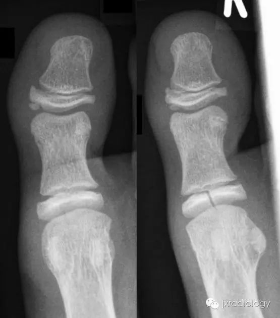
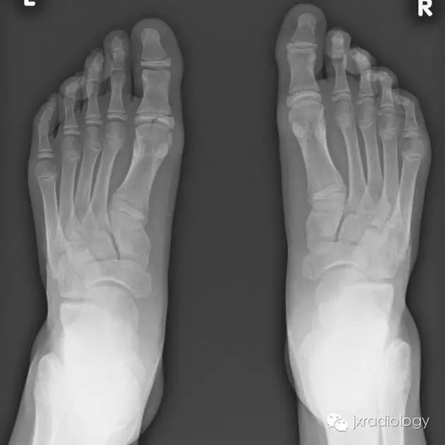
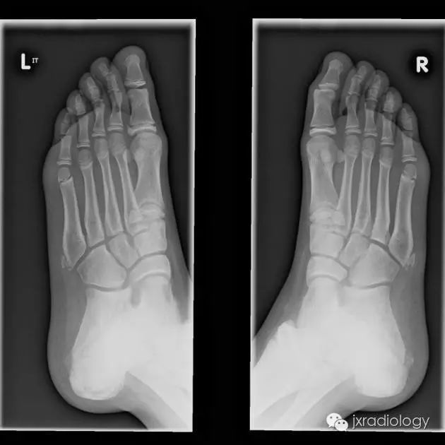
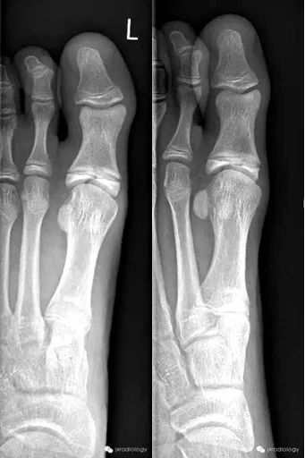
骨骺裂是一种正常变异。它可以是单侧或双侧,最常见的部位是足的第1趾近节趾骨的骨骺。
Cleft epiphysis is a normal variant of an epiphysis. It can be either unilateral or bilateral The most common site is the prepiphysis of the first proximal phalanx of the foot.
X线平片显示骨骺见透亮状裂隙影;透亮影的边缘是可变的,可能是锐利的或不规则的。骨骺裂可保留至生长板的融合。
Radiographic features
Plain radiograph
Plain radiographs will demonstrate a lucent defect in the epiphysis. The borders of the lucency are variable and may be sharp or irregular. The cleft remains till the fusion of growth plate.
鉴别诊断:
骨骺裂必须和骨折鉴别。一般骨折损伤2-3周后复查平片可见愈合的迹象。识别这个现象是重要的,以避免过度治疗和不必要的手术干预。
Differential diagnosis
A cleft epiphysis has to be differentiated from fracture. Generally fractures demonstrate signs of healing if the radiograph is repeated 2-3 weeks after injury. Recognition of this entity is important to avoid over treatment and unnecessary surgical intervention.
Cleft epiphysis意译为骨骺裂,结合临床病史及复查才能和骨折区分。
病例图片
骨骺裂
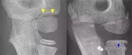
黄箭骨折;蓝箭是骨骺裂。LEFT a subtle lateral condyle fracture. Less than 2 mm displacement and probably stable. RIGHT a different case. Oblique view gives nice impression of fracture. Blue arrow indicates a cleft epiphysis of the radius (normal variant)