“不显影”髓内钉,让骨折复位和愈合评估更简单!
时间:2021-10-28 10:01:42 热度:37.1℃ 作者:网络
骨科内固定金属材料的特点为可显影,置入人体后可直接观察内植物位置是否准确,但对于术后评估来说,其可显影性可能会妨碍骨折愈合情况的观察。
碳纤维增强聚醚醚酮(carbon fiber-reinforced polyetheretherketone,CFR/PEEK)材料与金属内植物相比,不仅具有更好的抗疲劳特性,其最大的特点在于在影像上不显影,这一特性可能更利于骨折术后骨折部位愈合情况的观察。
对此,有学者对比粗隆间骨折,传统金属髓内钉与CFR/PEEK髓内钉在骨折随访过程中对骨折位置的评估及愈合进展的判断是否存在差异。

Introduction(介绍)
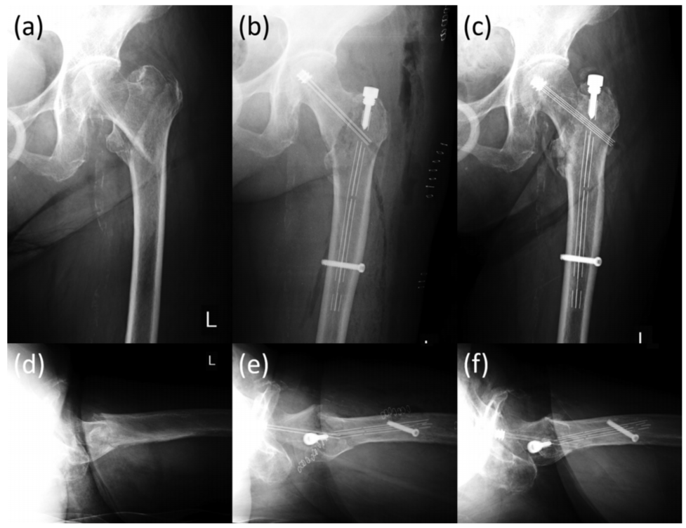
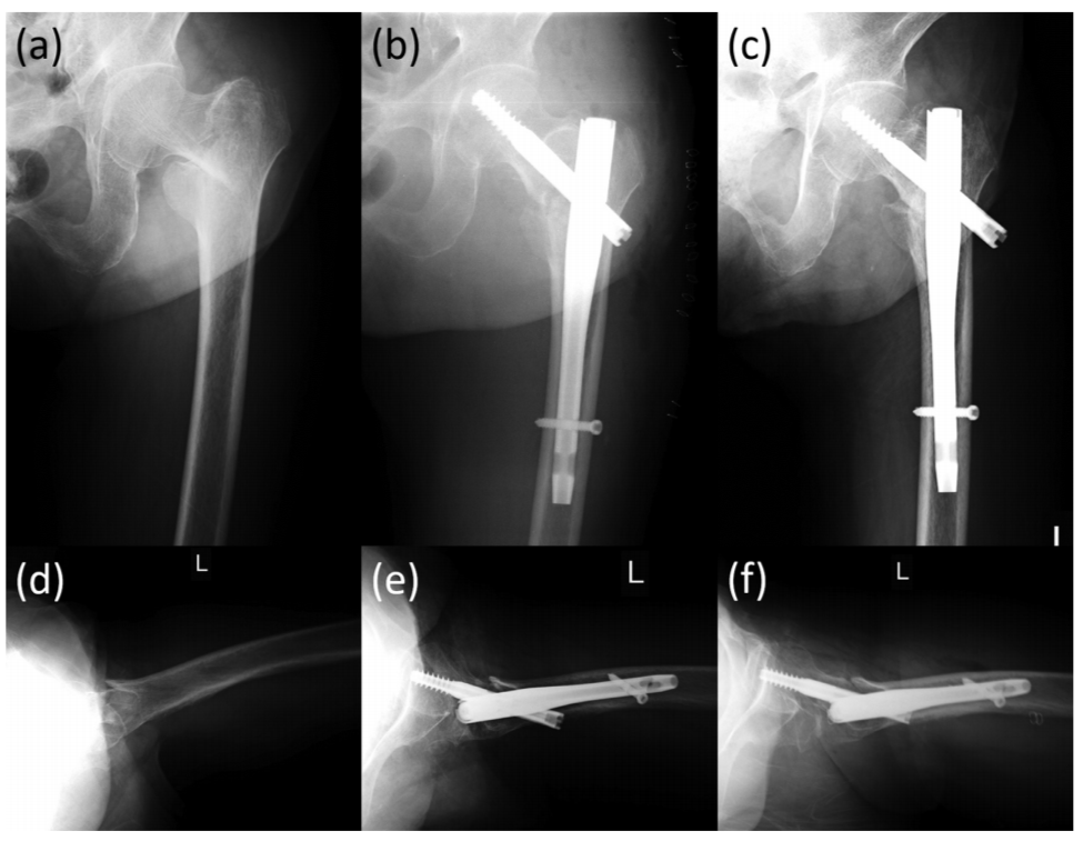
为了评估CFR/PEEK髓内钉因其可透过射线而在骨折愈合诊断中的优势,我们回顾性分析了使用CFR/PEEK髓内钉或传统金属髓内钉进行内固定的股骨粗隆间骨折的X线片和CT影像片。
[Introduction: To evaluate the advantages of a carbon fiber-reinforced polyetheretherketone (CFR/PEEK) intramedullary nail on the diagnosis of fracture healing because of its radiolucency, we retrospectively reviewed radiographs and computed tomography (CT) images of trochanteric femoral fractures that underwent internal fixation with the CFR/PEEK intramedullary nail or a traditional metallic intramedullary nail.]
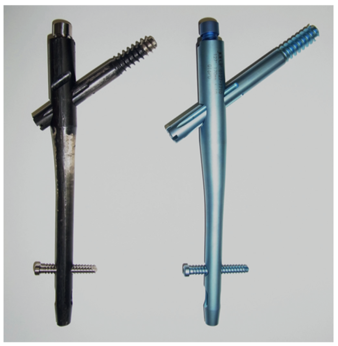
图1 CFR/PEEK髓内钉(左)与传统金属髓内钉(右)。
Methods(方法)
本文回顾了40例股骨粗隆间骨折患者的X线片和CT影像,其中20例患者接受了CFR/PEEK髓内钉治疗,20例患者接受了金属髓内钉治疗。在X线片的前后和侧位上将股骨粗隆间区域分成三个区域后,评估每个区域的骨折部位、骨折线和骨形成的可见性。在CT图像的三个轴向片上对散射的存在和散射对周围骨诊断的影响进行三级评估。
[Methods: Radiographs and CT images from 20 patients with intertrochanteric femoral fractures treated with a CFR/PEEK intramedullary nail and 20 similar patients treated with a metallic intramedullary nail were reviewed. After division of the intertrochanteric region into three zones on anteroposterior and lateral views of the radiographs, the visibilities of the fracture site, fracture line, and bone formation were evaluated in each zone. A three-grade assessment for existence of scattering and effect of scattering on diagnosis of the surrounding bone was performed on three axial slices of the CT images.]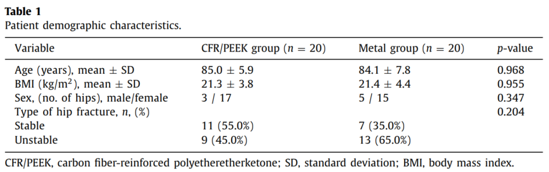
表1 两组患者一般资料比较。
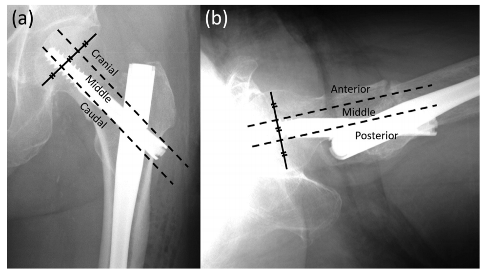
图2 在正位和侧位上对股骨粗隆部进行分区。正位上分为头区、中间区和尾区,侧位上分为前区、中间区和后区。
Results(结果)
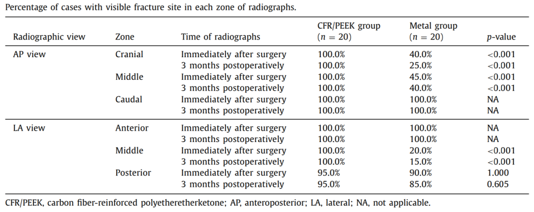
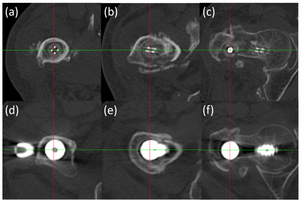
在CFR/PEEK组中,除了一个病例的侧位后区外,所有病例的所有区域均可见骨折部位。在X线的前后位上的头区和中间区以及侧位上的中间区,CFR/PEEK组的可见骨折部位率明显高于金属组。与金属组相比,CFR/PEEK组在CT图像上散射的存在等级和散射对周围骨诊断的影响显著降低。
[Results: In the CFR/PEEK group, the fracture site was visible in all zones for all cases except for the posterior zone on the lateral view in one case. In the cranial and middle zones on anteroposterior views and the middle zone on lateral views of the radiographs, the visible fracture site rates in the CFR/PEEK group were significantly higher than those in the metal group. The grades for existence of scattering and effect of scattering on diagnosis of surrounding bone on the CT images were significantly lower in the CFR/PEEK group compared with the metal group.]
Conclusion(结论)
与使用传统金属髓内钉治疗的病例相比,使用CFR/PEEK髓内钉治疗的病例在X光片上显示出优越的骨折部位可视性,从而证实了CFR/PEEK髓内钉在评估骨折复位和骨形成方面的优势。CFR/PEEK钉在CT图像上引起的散射很小,导致假体周围松质骨和皮质骨的诊断价值高于金属钉。
[Conclusion: Superior fracture site visibility on radiographs was demonstrated in cases treated with the CFR/PEEK intramedullary nail compared with cases treated with the traditional metallic intramedullary nail, thereby confirming the advantages of the CFR/PEEK intramedullary nail for evaluation of fracture reduction and bone formation. The CFR/PEEK nail evoked little scattering on CT images, leading to higher diagnostic values for the peri-prosthetic cancellous and cortical bone compared with the metallic nail.]